
Microscopic View Curd Seen Microscope Lactobacillus Stock Photo 1148034800 Shutterstock
Find Microscopic View Curd Seen Microscope Lactobacillus stock images in HD and millions of other royalty-free stock photos, illustrations and vectors in the Shutterstock collection. Thousands of new, high-quality pictures added every day.
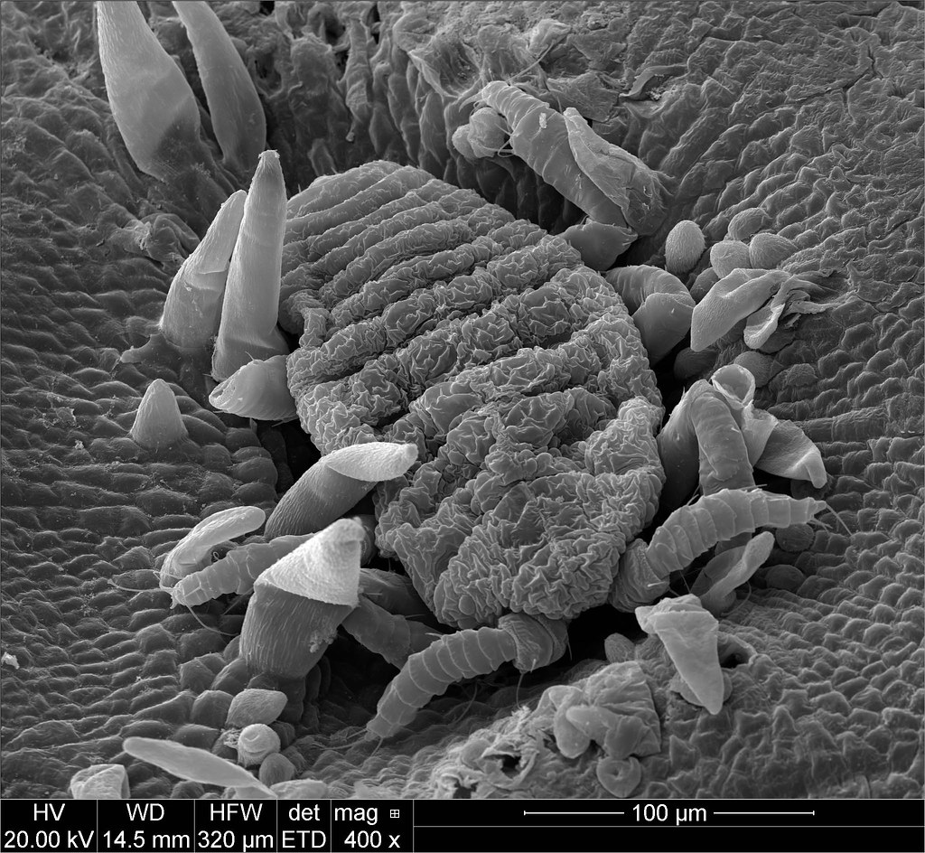
Microscopic view galls A microscopic view of a galling ins… Flickr
Old or expired curd microscopyAs you know, curd contains Lactobacillus (bacteria) and yeast cells (fungus) normally butthis old or expired curd having extra.

Microscopic View Curd Seen Microscope Lactobacillus Stock Photo 1148034749 Shutterstock
Remove excess solution around the coverslip with a paper towel or tissue. View in the compound microscope at 4 x or 10 x initially, before moving to higher magnification. Bacteria will appear small even at the highest magnification. NOTE: Step 2 is optional. You will be able to see the bacteria even without using the stain.

Side Effects of Eating Curd Everyday
The pressed curd or the cheese (477 ± 6 g) was removed from the mould and stored at 4 °C in a zip lock plastic bag from which the air had been removed. The gel and curd samples were analysed immediately after sampling, while the cheese samples were analysed within one week of pressing. 2.2. Cryo scanning electron microscopy

Microscopic View Curd Seen Microscope Lactobacillus Stock Photo 1148034803 Shutterstock
Curd microorganisms in gram stain are bacteria, Lactobacillus, and fungus, yeast cells as shown above picture. Curd is a dairy product and it is obtained by coagulating milk in a process called curdling. The coagulation can be achieved artificially by adding rennet or any edible acidic substance such as lemon juice or vinegar.

How curd looks under microscope yogurt under microscope // curd under microscope // Bacteria
View and focus specimens under a microscope. Determine total magnification of a specimen. Locate a specimen if given a slide. Introduction. In Biology, the compound light microscope is a useful tool for studying small specimens that are not visible to the naked eye. The microscope uses bright light to illuminate through the specimen and.

Microscope Yoghurt YouTube
SIM and iSIM both work off the concept of using structured patterns of light, with SIM making use of a Moiré pattern caused by the interference between two different patterns at an angle. iSIM imaging of live cell division with the Kinetix22 sCMOS. A) A C. elegans zygote, microtubules (green), and cell polarity marker PAR-2 (magneta).

Milk to Curd by Lactobacillus Bacteria Microscopic View YouTube
Knoop and Peters (46) studied the curd structures obtained by rennet and acid coagulation of milk using phase- contrast and electron microscopy. They found that the curd develops by chaining and clustering of casein micelles which gradually form a three-dimensional network. If the development of the network is interrupted, the casein precipitates.

What is Lactobacillus?
Anatomy of a Microscope - Field Curvature. Total internal reflection fluorescence microscopy (TIRFM) is an elegant optical technique utilized to observe single molecule fluorescence at surfaces and interfaces.. photomicrographers would often restrict the area recorded on film to the focused central area of the view field, thus obscuring the.

Microscopic View Curd Seen Microscope Lactobacillus Stock Photo 1148034755 Shutterstock
The LAB strains present in curd and vegetables are processed for pure culture isolation of bacteria. The presence of rod-shaped, gram-positive bacteria were confirmed by using gram staining and.

Microscopic View Curd Seen Microscope Lactobacillus Stock Photo 1148034764 Shutterstock
The human gut microbiota play a significant role in nutritional processes. The concept of probiotics has led to widespread consumption of food preparations containing probiotic microbes such as curd and yogurt. Curd prepared at home is consumed every day in most homes in southern India. In this study the home-made curd was evaluated for lactic.

Microscopic observation of Gram's stained Lactobacillus spp. Download Scientific Diagram
Electron Microscopic Studies of Casein Micelles and Curd Microstructure in Cottage Cheese. In cheese curd the same phenom- 1980 J Dairy Sci 63:37-48 37 38 GLASER ET AL. enon occurs, and in ripened cheeses such as Camembert and Cheddar the micelles agglo- merate to such an extent that their individual identity is lost (10, 11, 12.

Why Is Visualization Not Sufficient To Properly Identify Bacteria
View under the microscope starting with low power (for high power, add immersion oil) Observation (Discussion) Depending on the sample under investigation, students will have the opportunity to observe and identify the size and shape of the bacteria. They are categorized according to their shape (Morphology) and the how they stain (gram.

Microscopic image of a broccoli curd harvested on August 4 th , 2011... Download Scientific
Microscopic view of the Curd seen from the microscope. The Lactobacillus Bacteria. The Science education. Macro. Close up.

Microscopic View Curd Seen Microscope Lactobacillus Stock Photo 1148034782 Shutterstock
Hello dosto Aaj Hum Dahi Ko Microscope me dekhenge. ky sach me dahi me bacteia hotehai, kya wo bacteria hamari sehat ke liye acche hai ya bure. The species o.

Bacteria stock photo. Image of curd, microscope, bacteria 52660594
Microscopic Morphology. Gram staining slides were observed under microscope oil immersion. Endospore Test. Bacterial smear was made on microscopic slide under aseptic conditions and heat fixed. Then slide was placed over the steaming water bath and malachite green (primary stain) was applied for 5 minutes.